Dental pulp regeneration · matter, tissue, innovation
Regenerating the Living Tooth.
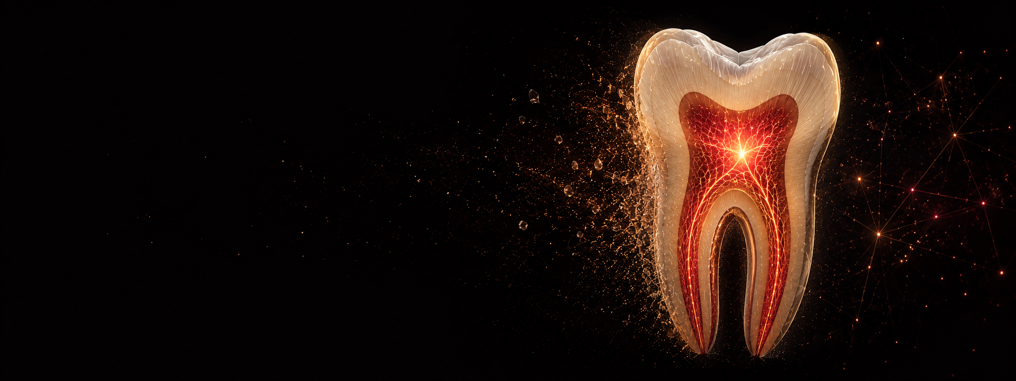
The dental pulp is the living tissue at the centre of the tooth, supporting sensation, defence, dentin formation and root development. Damage to this tissue can compromise both its biological function and the long-term behaviour of the tooth.
Matter for Vitality examines how bioactive materials, tissue engineering and emerging experimental approaches can support the preservation, study and reconstruction of viable pulp-like tissue within root anatomy.
Matter. Tissue. Innovation.
Three connected lines of work organise the programme.
How material chemistry, structure and ion release shape the local wound environment.
How cells, extracellular matrix and vascularisation organise within the confined anatomy of the root canal.
How emerging cell sources, experimental models and computational methods extend what can be tested and designed.
The material-tissue interface.
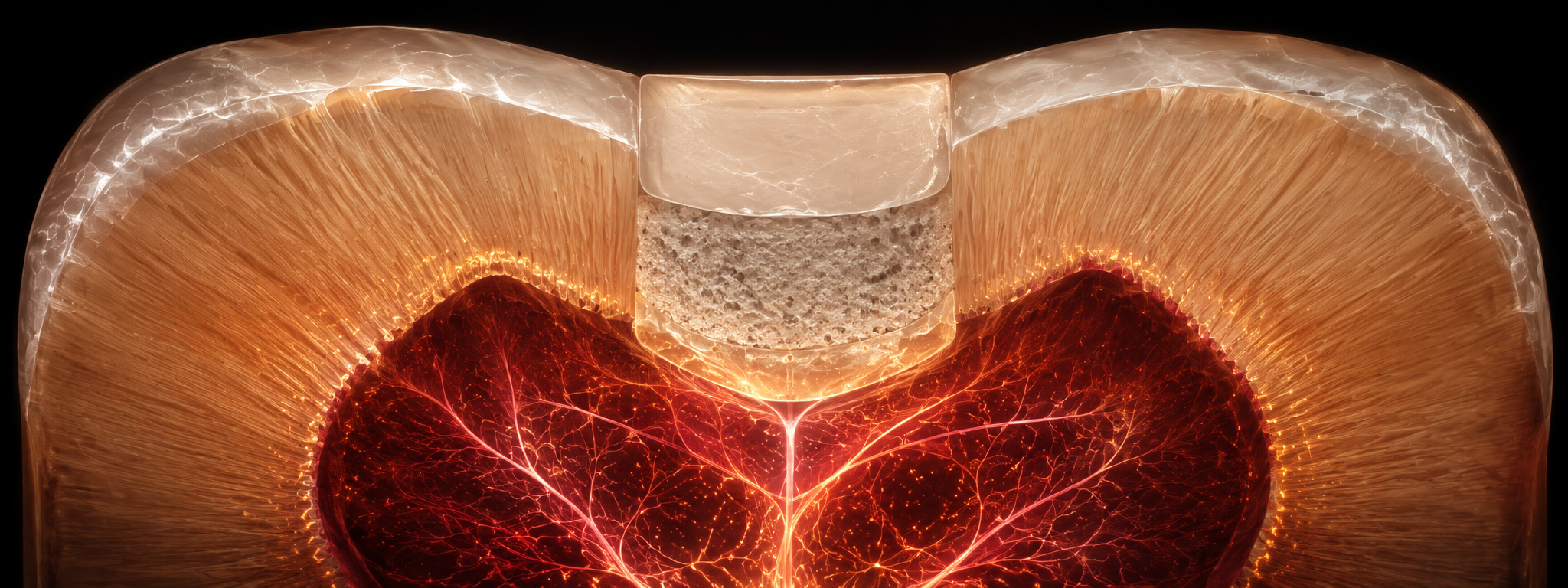
A calcium-silicate cement over exposed pulp, with a dentin bridge forming beneath it.
Materials placed close to the dental pulp must satisfy both physical and biological requirements. They need to seal the site, remain stable under clinical conditions and provide handling, setting, radiopacity and mechanical properties appropriate to their use.
At the exposed or injured interface, composition, surface characteristics, alkalinity and ion release also alter the local environment encountered by cells. These material-dependent conditions can influence cell survival, differentiation and mineral deposition, making the interface itself part of the biological response.
Evidence · Material formulation and biological response →Organising living tissue within root anatomy.
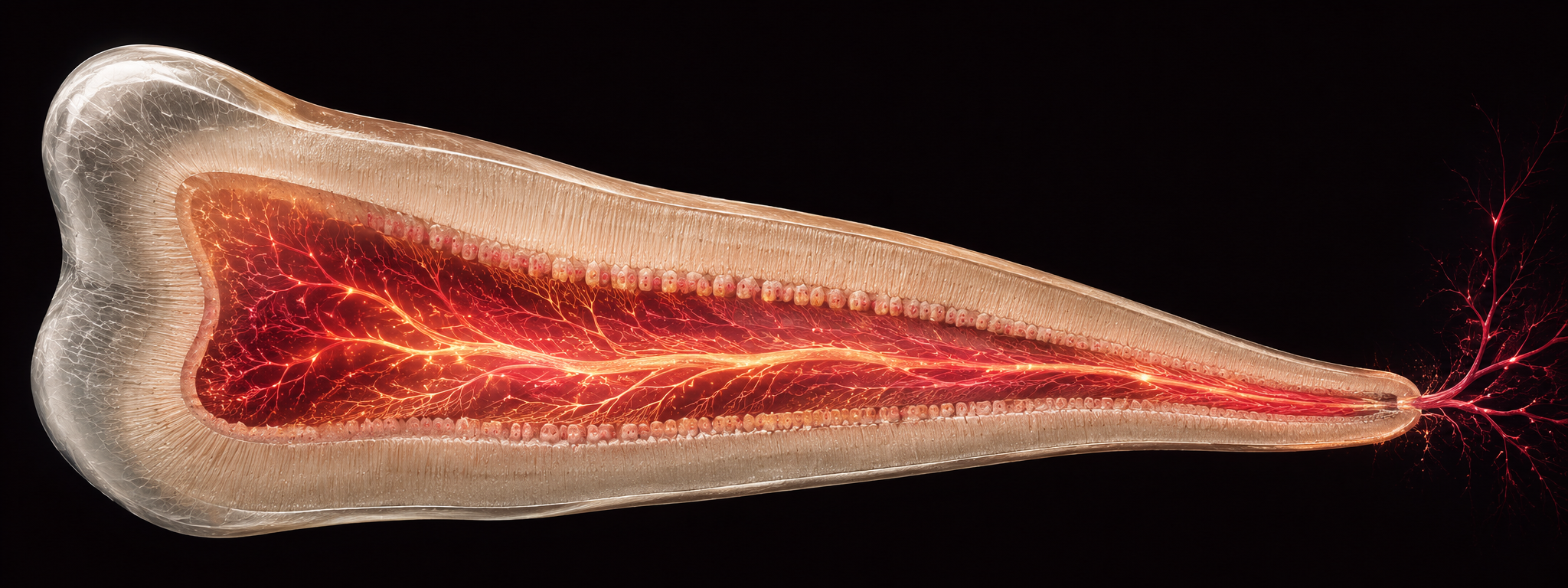
Conceptual illustration of vascularised pulp-like tissue occupying the root canal.
Pulp regeneration requires viable tissue to organise throughout the confined anatomy of the root canal while maintaining access to nutrients and vascular support. Experimental models have shown that cell-containing scaffolds can support pulp-like tissue formation and dentin-associated structures within full-length human roots. The continuing challenge is to reproduce this organisation under conditions that are biologically and clinically relevant.
Tissue organisation
Across the full canal
Vascularised pulp-like tissue and tubular dentin along full-length root canals.
Cell source
Toward odontoblast-like cells
A renewable experimental cell source capable of forming pulp-like tissue and tubular dentin.
Pulp tissue engineering
Complementary questions in tissue organisation and cell source.
These approaches were investigated in distinct experimental models.
Explore the pulp-regeneration researchConceptual model
Tissue in formation.
This sequence illustrates the biological requirements of pulp tissue engineering. It does not represent an established clinical procedure.
Scroll ↓ follow tissue formation through the canal
Space


Full-length human root-canal model
Human premolar roots containing SHED delivered in hydrogels developed vascularised pulp-like tissue and tubular dentin in vivo.
The model allowed tissue organisation, vascularisation and dentin-associated structures to be examined along the full length of the root.
View J Dent Res 2013
Extending the experimental toolkit.
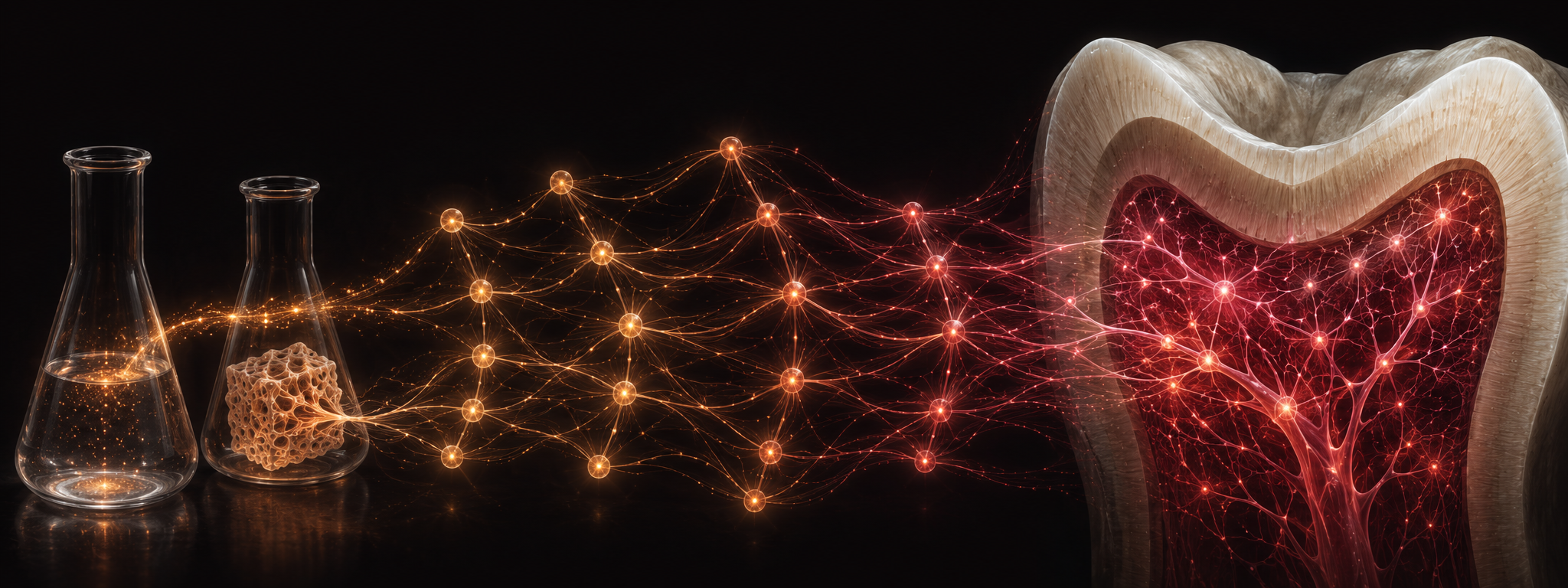
Illustration representing complementary biological and computational approaches within the programme.
The programme also examines approaches that extend what can be studied experimentally. On the biological side, this includes alternative cell sources and models for generating dentin-forming phenotypes. On the computational side, material datasets can be used to identify influential variables, predict outcomes and prioritise candidate formulations for experimental testing.
These approaches are complementary rather than interchangeable. Biological models address how cells and tissues form and function, while computational methods help navigate complex material design spaces.
How experimental models shape the evidence.

Illustration: different models and assays make different aspects of the same biology observable.
Every experimental model captures some aspects of pulp biology and omits others. Cell culture, dentin-based models and full-root systems differ in anatomical context, cell organisation, transport conditions and the biological functions they can reproduce.
Assays also have limits. Materials can interfere with measurements, and a positive result in one endpoint does not necessarily predict performance in another. Interpreting a study therefore requires attention to both the biological question and the model used to answer it.
Methodological guidance · Dental Materials 2025 →Published work supporting the programme.
The studies below trace the development of the programme across biomaterials, tissue models, cell sources and data-guided design.
Dental Pulp Tissue Engineering in Full-Length Human Root Canals
Demonstrated vascularised pulp-like tissue and tubular dentin formation within full-length human root canals.
Bioactivity, Physical and Chemical Properties of MTA Mixed with Propylene Glycol
Showed how a formulation change could improve flowability and calcium release without automatically improving cellular bioactivity.
Functional Odontoblastic-Like Cells Derived from Human iPSCs
Established a renewable experimental route to odontoblast-like cells capable of forming pulp-like tissue and tubular dentin in a subcutaneous dentin-disc model.
Taguchi’s Methods to Optimize the Properties and Bioactivity of 3D-Printed Polycaprolactone/Mineral Trioxide Aggregate Scaffolds
Used Taguchi design to identify which scaffold variables controlled pH, degradation, porosity and mechanical behaviour, then confirmed the predictions experimentally.
A Critical Analysis of Research Methods and Biological Experimental Models to Study Pulp Regeneration
Mapped how cell, tissue and animal models shape the questions, strengths and limitations of pulp-regeneration research.
Regenerating the Dental Pulp — Scaffold Materials and Approaches
Connected cell-based, cell-homing and scaffold strategies to the biological requirements of pulp tissue formation.
Designing Calcium Silicate Cements with On-Demand Properties for Precision Endodontics
Demonstrated target-driven, multi-property prediction of calcium-silicate cement recipes followed by experimental validation.
Guidance for Evaluating Biomaterials’ Properties and Biological Potential for Dental Pulp Tissue Engineering and Regeneration Research
Connected material characterisation, biological models, functional outcomes and reporting quality within a unified evaluation framework.
pH Prediction in Commercial and Experimental Calcium Silicate Cements via Material Informatics
Predicted long-term alkalinity from early pH measurements and specimen surface area, then validated the model across commercial and experimental calcium-silicate cements.